Mechanism of membrane perforation in rotavirus cell entry

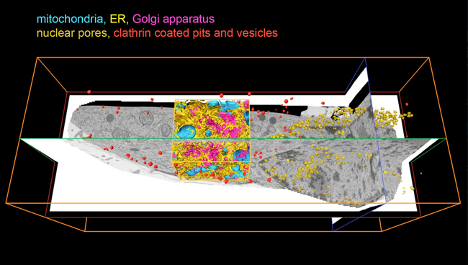
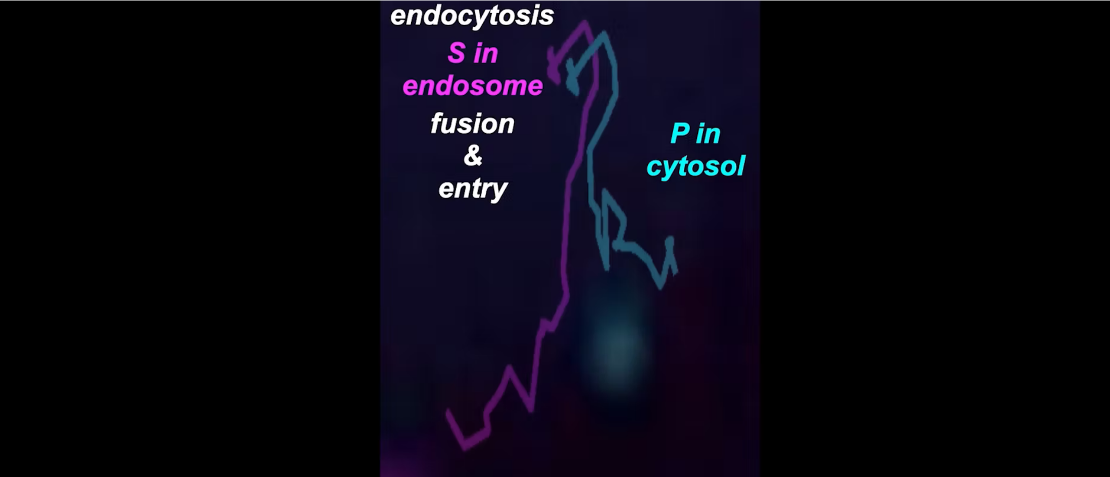
Cell entry of nonenveloped animal viruses requires translocation of a macromolecular assembly across a cellular membrane. Double-stranded RNA viruses introduce into the target cell an inner capsid particle that does not uncoat further. Instead, it extrudes capped viral mRNA by virtue of polymerase and capping activities within it. As described here, we used cryogenic electron tomography to visualize the full course of rhesus rotavirus entry, from cell attachment and virion uptake to release of the subviral particle.